Draw And Label Sponges - View The Images Of Sponges Having Observed Chegg Com : Label as many parts as you can see.
byAdmin•
0
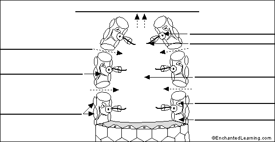
Draw And Label Sponges - View The Images Of Sponges Having Observed Chegg Com : Label as many parts as you can see.. Begin by drawing a large, curved square. In this video i'm going to draw diagram of sycon sponge labelled diagram easily and step by step and do it's classification in phylum porifera , classificati. A small clone grows on the side, falls off and develops into a new individual. The difference between this and the mechanisms of other animals. The sponge to draw in ambient water through small inhalant pores (ostia) and ilter out microscopic food particles.
Explain how a stinging cell in a cnidarian fires. The instructions included specific information about sponge anatomy to be colored on diagram. An ectoderm, or outer layer, and an endoderm, or inner layer. The primitive structure of a sponge consists of only two layers of cells separated by a. The food particles are caught by the collar of the choanocyte and brought into the cell by phagocytosis.
Label Sponge Cross Section Enchantedlearning Com from enchantedlearning.com List 4 ways sponges defend themselves. 124 s.if mature sponges are sessile, and bods just fall off, how can sponges be dispersed through the ocean? And the simple act of parts of a sponge breaking of and establishing in a new location. Meaning 'pore bearer'), are a basal animal clade as a sister of the diploblasts. Structure and function in sponges. Label sponge external anatomy diagram using the definitions listed below, label the sponge and the flow of water through it. Place the sample on a microscope slide, and a few drops of water and cover it with a coverslip. The growth of stolons that develop into new individuals;
The difference between this and the mechanisms of other animals.
A small clone grows on the side, falls off and develops into a new individual. The instructions included specific information about sponge anatomy to be colored on diagram. Porifera label this diagram of the sponge and identify the following osculum, estium, collar cells spicule, epidermis cells. Discover (and save!) your own pins on pinterest Label sponge external anatomy diagram using the definitions listed below, label the sponge and the flow of water through it. Where does a cell membrane come in contact with water. Sponges can reproduce in a variety of ways, both asexually and sexually. 4a) label the parts of a simple sponge and draw an arrow showing the flow of water throogh the sponge on figure 4. Observe the sponge under the compound scope. How to draw hydrahow to draw hydra, how to draw hydraulic circuit diagram, how to draw hydra step by step, how to draw hydra easy, how to draw hydra in zoolo. Drawings can highlight the important features of a specimen. To the right of the square, draw a curved rectangle to create a three dimensional effect. Sponges exhibit less specialization (adaptation of a cell for a particular function) of cells than most invertebrates.
Sponges exhibit less specialization (adaptation of a cell for a particular function) of cells than most invertebrates. Compare darwin's theory of evolution vs lamarck's theory. Be sure to use the following terms in your description: Drawings can highlight the important features of a specimen. Draw and label a cross section of a typical sponge.
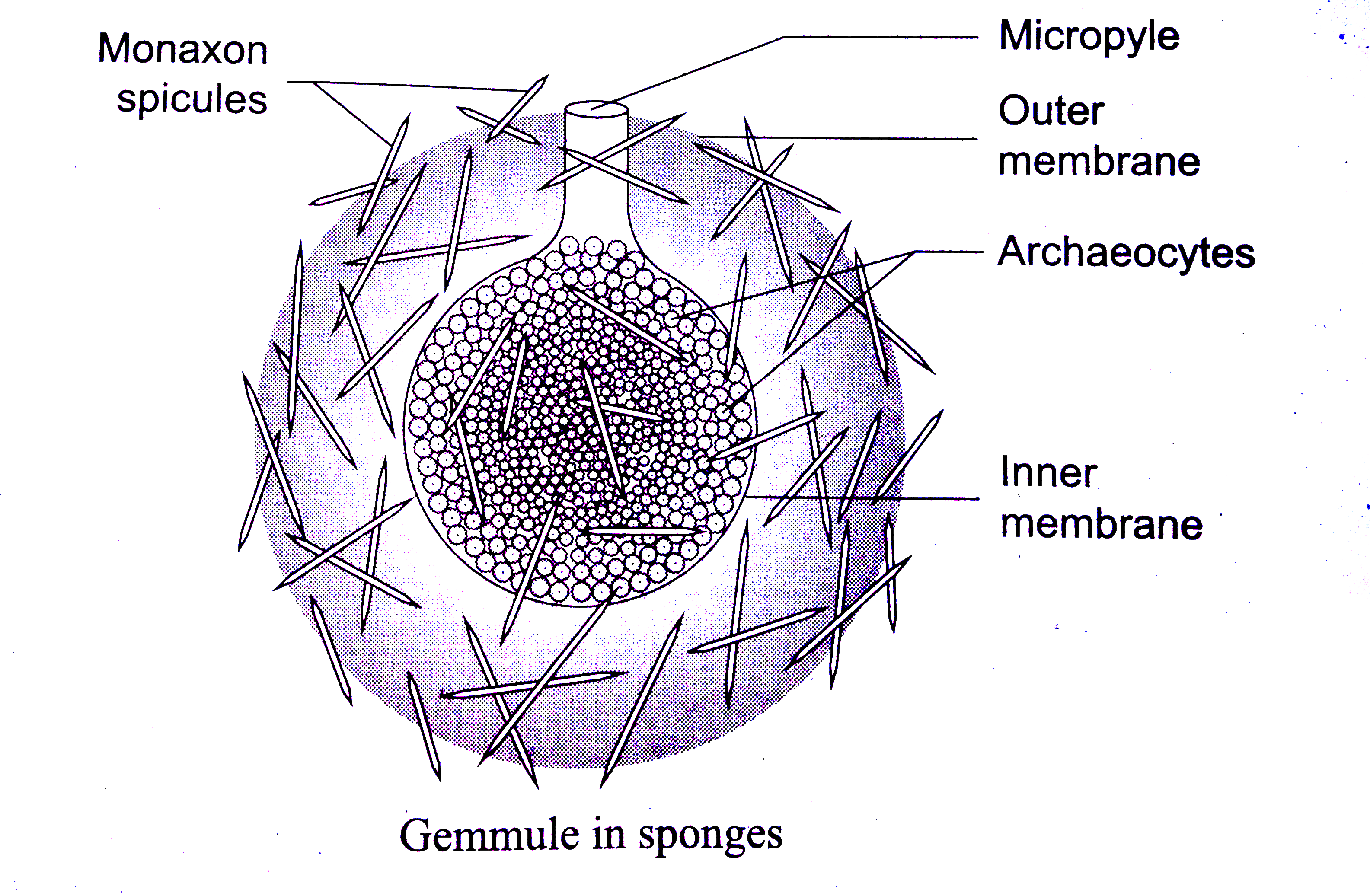
Draw And Label A Gemmule Of Sponge from d10lpgp6xz60nq.cloudfront.net The specialized cell types in sponges (b) each perform a distinct function. The sponges clipart gallery includes 48 illustrations of sponges. 4a) label the parts of a simple sponge and draw an arrow showing the flow of water throogh the sponge on figure 4. Filtered water is then expelled through fewer, larger exhalant openings (oscules). Sponges a coloring worksheet answer key original document: Make an illustration of your observations. Explain the process of reproduction in the sponge: The growth of stolons that develop into new individuals;
Be sure to use the following terms in your description:
The sponge to draw in ambient water through small inhalant pores (ostia) and ilter out microscopic food particles. Hermaphroditic, gamete, spawn, planktonic larvae. Be sure to use the following terms in your description: And the simple act of parts of a sponge breaking of and establishing in a new location. Digestion of the food particle takes place inside the cell. Hermaphroditic, gamete, spawn, planktonic larvae. Find a large selection of woodworking chisels including mortise chisels, corner chisels, draw knives and more at rockler. A bud separating from the parent sponge and creating a new sponge elsewhere; The sponges clipart gallery includes 48 illustrations of sponges. Drawing is a very important skill in biology and is considered a type of data collection because drawings help to record data from specimens. Using a hand lens.observe and draw the sponge. The primitive structure of a sponge consists of only two layers of cells separated by a. Be sure to use the following terms in your description:
Where does a cell membrane come in contact with water. Sponges, also called poriferans, are in the phylum porifera and are all sessile animals that live and feed attached to the bottom of the sea. Membranes 241 draw and label a diagram to show the structure of membranes. Filtered water is then expelled through fewer, larger exhalant openings (oscules). Label the bottles in the presence of the patient with either cerner or epic labels.
Sponge Structure And Function Advanced Read Biology Ck 12 Foundation from dr282zn36sxxg.cloudfront.net Explain how a stinging cell in a cnidarian fires. Filtered water is then expelled through fewer, larger exhalant openings (oscules). 31 sketch and label a phospholipid due aug 31 2016 by 1159pm. Record the time, collection site, and your initials on the requisition. An ectoderm, or outer layer, and an endoderm, or inner layer. Use 31 the cell membrane as a reference. Structure and function in sponges. The difference between this and the mechanisms of other animals.
Label as many parts as you can see.
The food particles are caught by the collar of the choanocyte and brought into the cell by phagocytosis. Where does a cell membrane come in contact with water. Place the sample on a microscope slide, and a few drops of water and cover it with a coverslip. Wandering, pseudopod bearing cells in the mesohyl; Draw and label a cross section of a typical sponge. Hermaphroditic, gamete, spawn, planktonic larvae. Hermaphroditic, gamete, spawn, planktonic larvae. The sponge's (a) basic body plan is a cylinder shape with a large central cavity. Discover (and save!) your own pins on pinterest Sponges are diploblasts meaning that they develop from two basic germ layers: Sponges, the members of the phylum porifera (/ p ə ˈ r ɪ f ər ə /; Be sure to use the following terms in your description: Drawings can highlight the important features of a specimen.